

Corals Revealed as Never Before Through a Groundbreaking New Microscope in California
A groundbreaking underwater microscope developed at Scripps Institution of Oceanography is giving scientists their first up-close look at coral stress before bleaching begins.
Updated
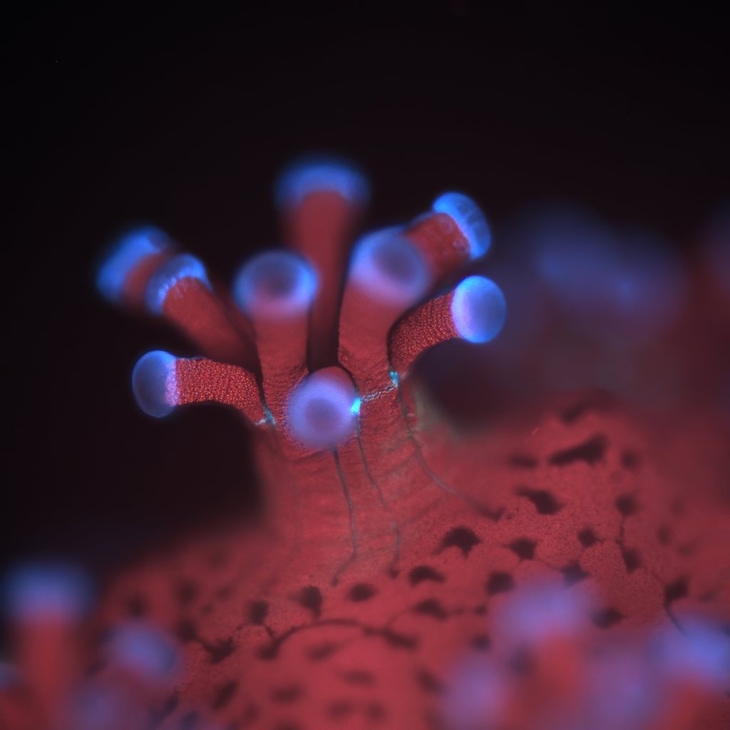 A fluorescence image of a polyp from the coral_ Stylophora pistillata_ (side view) taken with the BUMP. (Credit: Or Ben-Zvi)
(We did a video about this story as well. We hope you watch!)
The story of corals in the modern age on this planet is one of near-total despair. I’ve done several stories on corals and have spent many hours diving reefs around the world, from the Mesoamerican Reef in Belize to the unbelievably robust and dazzling reefs in Indonesia. There are still some incredible places where corals survive, but they are becoming fewer and farther between. I don’t want to get too deep into all the statistics, but suffice it to say: scientists estimate that we have already lost about half of the world’s corals since the 1950s, and that number could rise to as much as 90 percent by 2050 if current rates of bleaching and die-offs continue.
What’s crazy is that we still don’t completely understand corals, or exactly why they are dying. We know that corals are symbionts with microscopic algae called zooxanthellae (pronounced zo-zan-THEL-ee). The corals provide cover, a place to live, and nutrients for the algae. In return, the algae provide sugars and oxygen through photosynthesis, fueling coral growth and reef-building. But when the planet warms, or when waters become too acidic, the relationship often collapses. The algae either die or flee the coral. Without that steady food source—what one scientist I interviewed for this story called “a candy store”—corals turn ghostly white in a process known as bleaching. If stressful conditions persist, they starve and die.
But why?
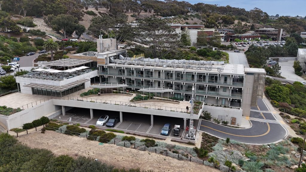 Scripps Institution of Oceanography in La Jolla (Photo: Erik Olsen)
“We still have no idea, physiologically, in the types of environments where bleaching predominates, whether the animal is throwing them out because it’s going to try to survive, or whether the little tiny plants say to the animal, ‘look, we can’t get along in this environment, so we got to go somewhere else’” says Dr. Jules Jaffe, an oceanographer at the Scripps Institution of Oceanography at the University of California, San Diego in La Jolla, California, and the head of the Jaffe Laboratory for Underwater Imaging.
The Great Barrier Reef, once Earth’s largest living structure, has suffered repeated mass bleaching events since 1998, several of them in just the last decade, and big stretches of it are now graveyards of coral skeletons. Scientists around the world are racing to figure out what’s going on, and how to at least slow down the bleaching that’s hitting nearly every major reef system.
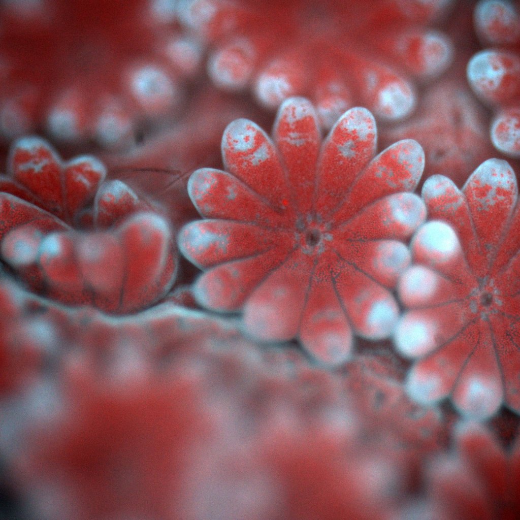 An image of Montipora coral polyps taken with the BUMP. Each polyp has a mouth and a set of tentacles and the red dots are individual microalgae residing inside the coral tissue. (Photo: Or Ben-Zvi)
One place where scientists are making small strides is at the Jaffe Lab, which I visited with my colleague Tod Mesirow and where researchers like Dr. Jaffe and Dr. Or Ben-Zvi have developed a new kind of underwater microscope that allows them to get close enough to corals to actually see the algae in action.
That’s not easy. Zooxanthellae are only about 5 to 10 microns across, about one-tenth the width of a human hair, and invisible to the naked eye. With the new microscope and camera, though, you can see them in amazing detail. The lab has filmed things nobody had seen before, including corals fighting with each other for space, fusing together, and even responding to invading algae.
When I first reported on this imaging system years ago, it was still in its early stages. At the time, it was known as the BUM for Benthic Underwater Microscope. Since then, the Scripps team has added a powerful new capability: a pulsing blue light that lets them measure photosynthesis in real time. They call it pulse amplitude modulated light or PAM, and so now the system is known as the BUMP.
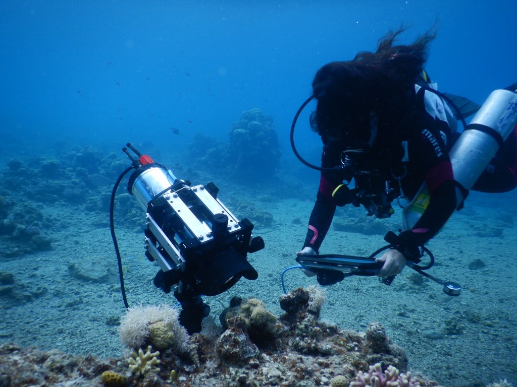 A field deployment of the BUMP in the Red Sea, where local corals were imaged and measured. (Photo: Or Ben-Zvi)
Here’s how it works: blue excitation light stimulates the algae’s chlorophyll, which then re-emits some of that energy as red fluorescence. By tracking how much of this red fluorescence is produced, researchers can calculate indices of photochemical efficiency, essentially how well the algae are converting light into energy for photosynthesis. This doesn’t give a direct count of sugars or photons consumed, but it does provide a reliable window into the health and productivity of the algae, and by extension, the coral itself.
And here’s the key part: all of this happens in the ocean, on living reefs, not in an aquarium or a lab.
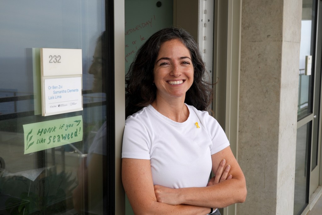 Dr. Or Ben-Zvi, researcher at Scripps Institution of Oceanography (Photo: Erik Olsen)
You can’t solve a problem you can’t see, and places like Scripps are where scientists build the tools that let us see the ocean in new ways. “There’s so much to learn about the ocean and its ecosystems, and my own key to understanding them is really the development of new instrumentation,” says Jaffe.
Dr. Ben-Zvi gave us a demonstration of how the system works in an aquarium holding several species of corals, including Stylophora, a common collector’s coral. The camera-microscope lit up and brought into crisp focus the tiny coral polyps along with their algal partners. On the screen we watched them in real time, tentacles waving as they absorbed the flashes of light from the BUMP, appearing, almost, as if they were dancing happily.
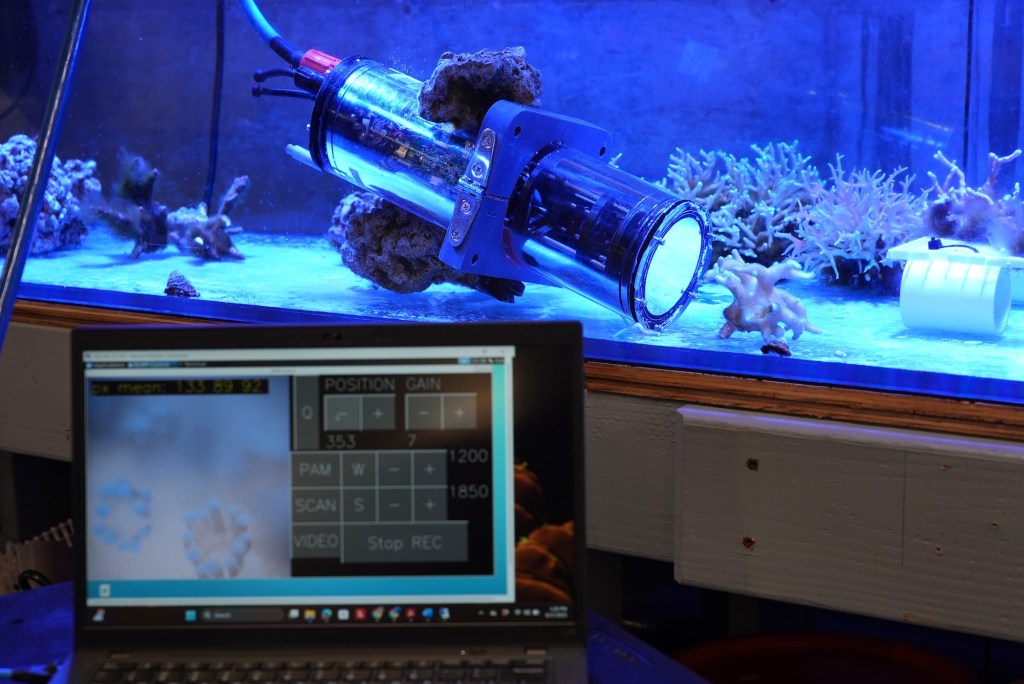 The Benthic Underwater Microscope PAM (BUMP) in action in the lab at Scripps Institution of Oceanography in La Jolla.
(Photo: Erik Olsen)
The Benthic Underwater Microscope PAM (BUMP) in action in the lab at Scripps Institution of Oceanography in La Jolla.
(Photo: Erik Olsen)
What this new tool allows scientists to do is determine whether corals may be under stress from factors like warming seas, pollution, or disease. Ideally, these warning signs are detected before the corals expel their zooxanthellae and bleach. Researchers are also learning far more about the everyday behavior of corals: something rarely studied in situ, directly in the ocean.
That in-their-native-environment aspect of the work is crucial, because corals often behave very differently in aquariums than they do on wild reefs. That’s where this microscope promises to be a powerful tool: offering insights into how corals really live, fight, and respond to stress.
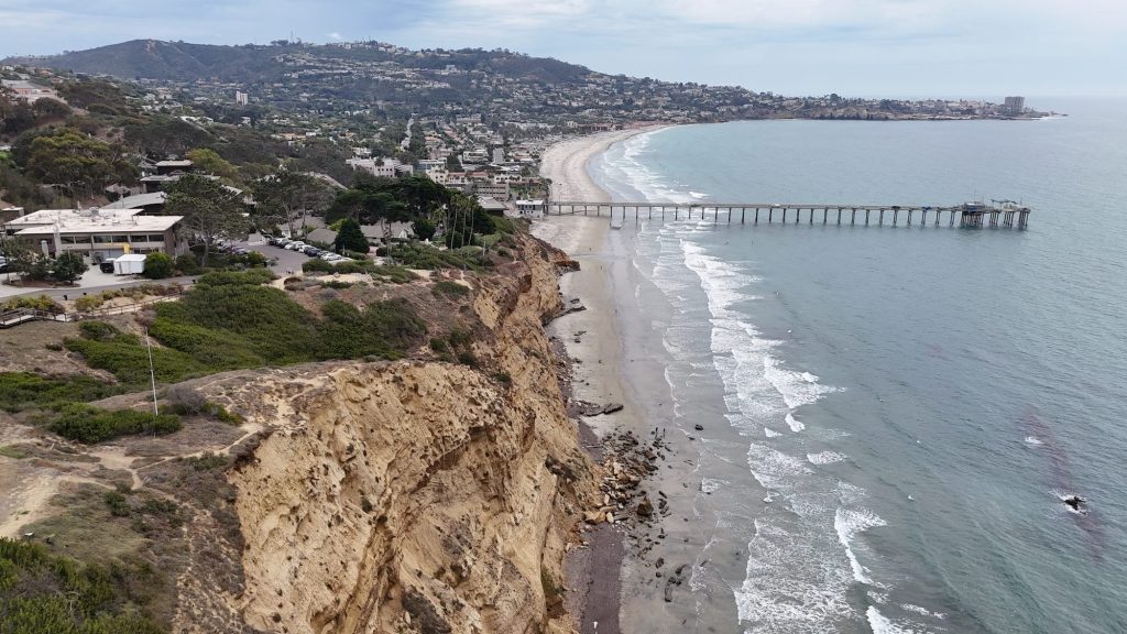 The view of La Jolla from the Scripps Institution of Oceanography (Photo: Erik Olsen)
Of course, what we do once we document a reef under stress is another matter. Dr. Ben-Zvi suggests there may be possibilities for remediation, though she admits it’s difficult to know exactly what those are. Perhaps reducing pollution, limiting fishing, or cutting ship traffic in vulnerable areas could help. But given that we seem unable—or unwilling—to stop the warming of the seas, these measures can feel like stopgaps rather than solutions. Still, knowledge is the foundation for any action, and this new tool is a breakthrough for coral imaging. If deployed widely, it could generate an invaluable dataset for researchers around the globe. The scientists behind it even hope to build multiple systems, perhaps commercializing them, to vastly expand the reach of this kind of monitoring.
But even Jaffe concedes it may already be too late: “Could a world exist without corals? Yeah, I think so,” he said. “It would be sad, but it’s going that way.”
Still, the images are stunning, and at the very least they might jolt people into realizing this is a crisis worth fighting. If we can’t fix it, future generations may have only pictures like these to remember the reefs.
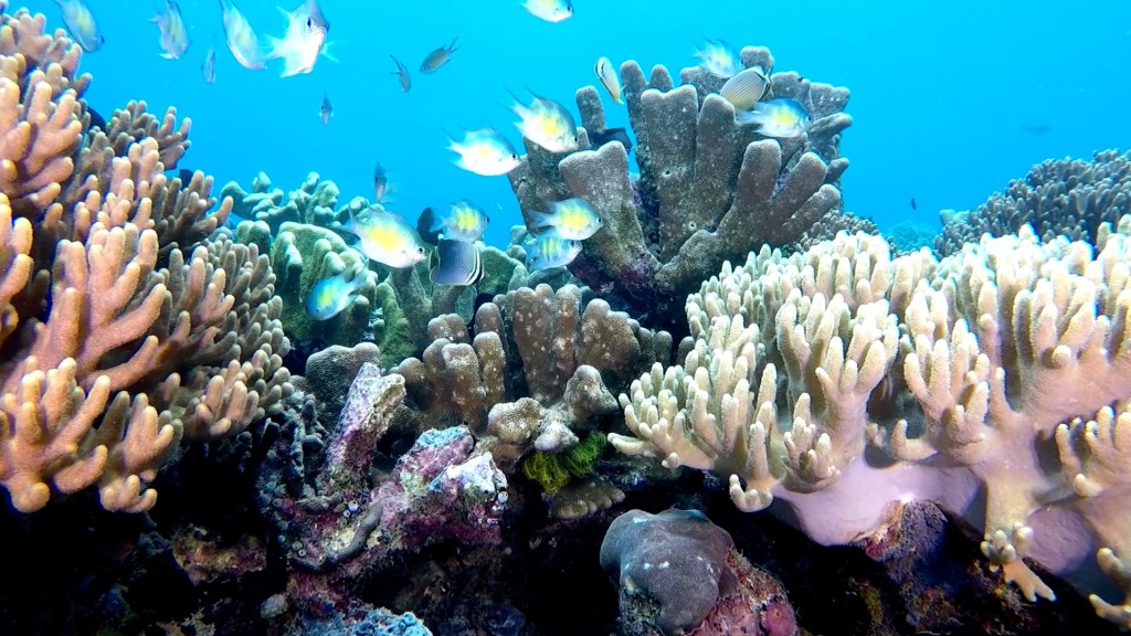 A healthy coral reef in Indonesia (Photo: Erik Olsen)
Is that valuable? Yes, but not nearly as valuable as saving the living reefs themselves. Dr. Jaffe told us,
“I’m on a mission to help people feel empathy toward the creatures of the sea. At the same time, we need to learn just how beautiful they are. For me, the combination of beauty and science has been at the heart of my life’s work.”
That’s really what this microscope is for. It’s a science tool, but it’s also a way to make people care about something they’d never otherwise see. Too bad the clock is ticking so fast.
(We did a video about this story as well. We hope you watch!)
More California science, photography and wild places on Instagram @californiacuratedus